
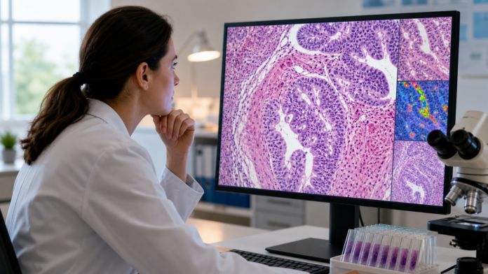
A new analysis of a major UK trial suggests routine biopsy slides may help identify which patients could benefit from a more intensive treatment.
A useful signal inside an existing trial
Researchers led by University College London have found a promising way to separate patients who appeared to benefit from an intensified treatment for locally advanced rectal cancer from those who did not. Their analysis used artificial intelligence to examine standard biopsy images collected before treatment.
The work revisited samples from the phase III ARISTOTLE trial, which enrolled patients through 75 hospitals in the United Kingdom. The original trial tested whether adding the chemotherapy drug irinotecan to standard chemoradiotherapy improved results before surgery. Across the trial population as a whole, adding irinotecan did not produce a general improvement.
The new study asked a more focused question: could a measurable feature inside the original tumour samples reveal a subgroup that responded differently? The researchers trained an image-analysis system to distinguish cancer cells from surrounding tissue and calculate tumour cell density. They then applied it to 414 diagnostic biopsy slides from trial participants.
That division produced two groups. The analysis classified 188 samples as having high tumour cell density and 226 as having low density. The treatment difference appeared in the high-density group, rather than across every patient.
Biopsy images become decision-making data
Among participants in the high-density group, adding irinotecan was associated with an approximately 43 percent lower risk of cancer recurrence and an approximately 50 percent lower risk of death over five years compared with standard chemoradiotherapy. The low-density group showed no comparable difference.
This is encouraging because the underlying material is already familiar to hospitals. Pathologists routinely examine stained tissue slides to diagnose cancer. The AI method is designed to count and classify millions of cells in digitized versions of those slides, turning a time-consuming measurement into something that could potentially be performed at scale.
The researchers have also developed a free online research tool called Octopath. Its purpose is to analyze uploaded biopsy-slide images, although the study team says the approach still needs further verification before it can guide routine treatment decisions.
The potential benefit is not simply finding more treatment. It is finding a better match between treatment intensity and the biology visible in an individual tumour. That distinction matters when an additional drug can also bring additional side effects.
A promising result with important limits
This was a post-hoc analysis, meaning the researchers looked back at trial material to test a new way of identifying responsive patients. It was not a new randomized trial in which treatment was assigned using the AI classification from the beginning.
For that reason, the result should not be read as a new standard of care or a tool ready for general clinical use. The UCL team explicitly says independent verification and additional clinical studies are required. The finding also applies to a defined group with locally advanced rectal cancer; it is not evidence about every stage or type of colorectal cancer.
The contrast with the original trial result is essential. Irinotecan did not improve outcomes for everyone. The hopeful finding is that tumour cell density may help reveal a subgroup for whom the balance of benefit and burden is different. That hypothesis now needs prospective testing.
Precision can also mean avoiding unnecessary strain
Cancer treatment decisions often involve weighing potential benefit against real physical costs. Irinotecan can intensify side effects when added to chemoradiotherapy. A reliable way to identify likely non-beneficiaries could therefore be valuable as well, because it might spare some patients an additional drug that is unlikely to help them.
The study illustrates a grounded use of medical AI: extracting more information from routine samples rather than replacing clinicians or promising an automatic cure. It also shows why broad trial results sometimes deserve carefully designed follow-up questions. A treatment that offers little average benefit may still matter for a biologically distinct subgroup.
The next step is confirmation, not celebration without limits. If independent studies reproduce the signal and prospective trials show that it can safely guide care, an ordinary biopsy slide could become a more useful map for personalizing a difficult treatment. For patients and clinicians, that would be meaningful progress toward giving intensive therapy to the people most likely to benefit from it.